How to read chest e-rays
working as an Intensiveness, ICU consultant
is actually based on the request from all of you guys, from my juniors from my colleagues
and this is all about reading and interpreting chest x-rays in a simplified manner and in this
video I have discussed around 25 x-rays and that too starting from the basics which you need to
know. So do watch this video till the end to become a chest x-ray expert. So let's begin
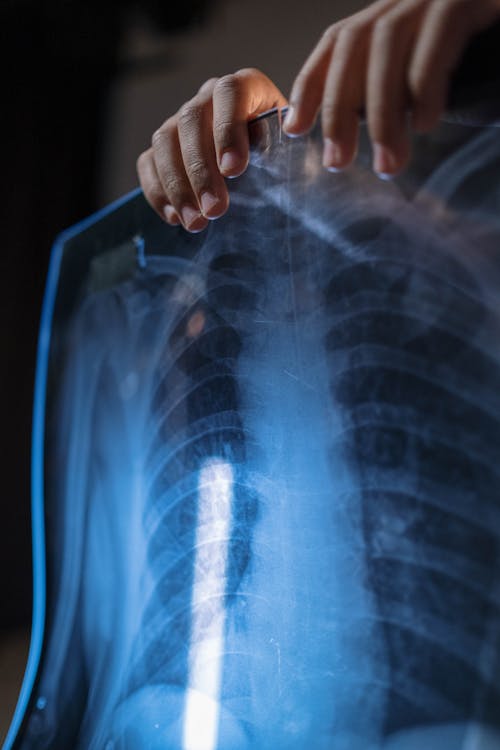
Before you start reading or interpreting the x-ray, you need to know which is the right
and the left side in this x-ray. This is the right side and this one is the
left side and this is usually marked on the x-rays. A good way to remember is that you
imagine that the patient is always facing towards you, this is true for both PA and AP films now
I will tell you the structures which are normally seen on chest x-ray. I'll just mark and name the
structures, will discuss in detail in the later part of this video. So this is the trachea,
this is the carina, right bronchus and this one is left bronchus this is divided into right and
the left bronchus then this is the lung field this one is the clavicle then this is aortic
knuckle this is the hilum over here is the heart this one is a diaphragm the right hemidiaphragm
these are the ribs and this one as you can see below the left hemidiaphragm is a gastric bubble
now this x-ray looks like a perfect x-ray right and before moving further you need to check the
patient detail this is very important and it is always mentioned on the x-ray
now the name is Chirag Madaan. Oh, I know this guy okay okay that's me . So check the name
the age of the patient the unique id number and the date on which it is taken next is
to assess the quality of the image it can be remembered by mnemonic RIPE where R stands for
rotation so as to see whether the film is rotated or not the spinous processes should lie halfway
between the medial ends of the clavicle that means in between the middle ends of
02:41
the clavicle if the patient is rotated then interpretation of the x-ray becomes difficult
02:48
firstly it may be difficult to know if the trachea is deviated to one side
by a disease process and it also becomes difficult to comment actually uh upon the heart size
and the changes in the lung density due to asymmetry of the overlying soft tissue
may be incorrectly interpreted as lung disease so now let's see what happens to the heart size
with respect to the rotation so this is a person standing over
and this is a normal the rays are coming from the back so this is most likely a pa film and
if the patient is rotated towards the left let's see the second second image. Second image shows
patient is rotated towards the left so as you can see the xray beams are going like this
and heart size looks greater or magnified or exaggerated whereas
if you see the third image the patient is rotated towards the right in this the true
size of the heart may be underestimated. Now next in RIPE is inspiration ideally
chest x-ray should be done in inspiratory phase if the x-ray taken during a good inspiratory effort
diaphragm should be intersected by five to six anterior ribs or eight to ten posterior
ribs in the mid clavicular line. Less is a sign of incomplete inspiration whereas if more than seven
anterior ribs then it suggests hyper inflation now let's count in this x-ray. So these are the
posterior ribs whereas these one are anterior one let's count the anterior ones this is first
second and third so there are three anterior ribs intersecting the diaphragm in mid clavicular area
that means this is either expiratory film or poor inspiratory effort also heart appears bigger and
there seems to be consolidation on left base now let's see the next x-ray so the number of anterior
ribs in this x-ray are you can calculate one this one is second then third then fourth fifth
and sixth heart seems to be normal and there is no consolidation at the base. now don't get
surprised my friends these two x-rays are of the same patient one in the expiratory
phase the first one and the second one in the inspiratory phase now why such a big difference,
first of all diaphragm pulls the under surface of the heart down during a good inspiratory effort
which is absent in expiratory phase so heart appears bigger in transverse diameter whereas
the second reason is the failure to distance the lungs fully can cause crowding the vessels


Comments
Post a Comment